Students can use NCERT Class 9 Advanced Science Solutions Chapter 9 Microscope and Microscopy Question Answer to understand complex concepts with ease.
Microscope and Microscopy Class 9 Questions and Answers
Microscope and Microscopy Question Answer Class 9
Let us Find Out
Question 1.
If a cell measures 5 mm on 100X image, calculate its actual size.
Answer:
Actual size = Image size/ Magnification = 5 mm/ 100 = 0.05 mm The actual size of the cell is 0.05 mm or 50 pm.
Question 2.
If you use a 15X eyepiece and 10X objective, what will be the total magnification?
Answer:
The total magnification (M) of a microscope is calculated by multiplying the magnification of the objective lens by that of the eyepiece lens:
M = m0 × me
= 10 × 15 = 150X
The total magnification is 150X.

Question 3.
If 4 cells fit across a 0.8 mm field of view, what will be the approximate size of one cell?
Answer:
Cell size = \(\frac{\text { Field of view }}{\text { Number of cells }}\)
= \(\frac{0.8 \mathrm{~mm}}{4}\) = 0.2 mm
The approximate size of one cell is 0.2 mm or 200 pm.
Think
Question 1.
Why do you think electron microscopes are usually found in big research centres and not in normal school laboratories?
Answer:
Electron microscopes are powerful but too costly, delicate, and advanced for school labs, so they are kept in big research centres and universities where proper facilities and experts are available.
Check Your Understanding
1. A microscope has a 10X eyepiece and a 40X objective.
(a) What is its total magnification?
(b) At this setting, the field of view is 0.4 mm. If 4 cells fit across, estimate the size of one cell.
Answer:
(a) Total Magnification = Eyepiece × Objective
= 10X × 40X = 400X
(b) Cell size = \(\frac{\text { Field of view }}{\text { Number of cells }}\) = \(\frac{0.4 \mathrm{~mm}}{4}\)
= 0.1 mm (or 100 μm)
Question 2.
(a) You want to watch live protozoa moving in pond water. Which microscope (light, phase-contrast, TEM, SEM) is best and why?
(b) Neha wants to study the 3D surface of a pollen grain. Which microscope should she choose and why?
Answer:
(a) Phase-Contrast Microscope.
Phase-contrast microscopy allows for the viewing of living cells in their natural, active state because it enhances contrast without the need for chemical stains, which would otherwise kill the specimen. While a standard light microscope can also observe living cells, phase-contrast provides a much clearer, high-contrast image of the moving organisms.
(b) Scanning Electron Microscope (SEM).
The SEM is specifically designed to give 3D-like images of surfaces by scanning the specimen with an electron beam. It is the standard tool used for detailed surface observations of objects like pollen grains, insect legs, and microchips.
Question 3.
Riya sees a sharp onion cell image at 100X, but when she switches to 400X, the image is big but very blurred. Name the concept causing this problem. Explain the reason.
Answer:
The concept responsible is resolution (resolving power).
Resolution is the ability to distinguish two close points as separate entities. If a microscope has a low resolution, increasing the magnification just creates “empty magnification”—the image becomes bigger, but the fine details are not captured, resulting in a blurry or fuzzy appearance. Therefore, magnification alone does not guarantee a better image. To observe fine details clearly, a microscope must have high resolution, which provides a sharper and more detailed image.
Question 4.
Draw a ray diagram of a compound microscope.
Answer:
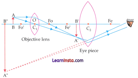
Question 5.
Design a simple poster “How to take care of a microscope?” with three do’s and three don’ts.
Answer:

Question 6.
At 40X total magnification, the field diameter is 4 mm. Predict the field diameter at 400X magnification (assume it is inversely proportional to magnification).
Answer:
Since the field diameter (D) is inversely proportional to the magnification (M):
M1 × D1 = M2 × D2
Given:
- Initial magnification (M1): 40X
- Initial Field Diameter (D1): 4 mm
- New Magnification (M2): 400X
- New Field Diameter (D2):D2
Calculation:
Plug the values into the formula:
40 × 4 = 400 × D2
160 = 400 × D2
D1 = \(\frac{160}{400}\)
D1 = 0.4 mm
Question 7.
A student accidentally traps many air bubbles while placing the cover slip. How will this affect observation? Suggest two ways to avoid bubbles next time.
Answer:
Effect on Observation:
- Air bubbles appear as dark circular structures.
- They scatter light and reduce image clarity.
- They may hide important specimen details.
Ways to avoid bubbles
- Lower the coverslip gently at an angle of about 45° to prevent air from getting trapped underneath.
- Ensure that a sufficient amount of mounting medium is present on the specimen before placing the coverslip.
- Place the coverslip slowly and carefully to allow the mounting medium to spread evenly beneath it.
- Check the slide for any trapped air bubbles and prepare the mount again if necessary. (Any two)
Question 8.
Compare TEM and SEM in terms of:
• Type of image.
• Best use (internal vs surface).
Answer:
The comparison between the Transmission Electron Microscope (TEM) and the Scanning Electron Microscope (SEM):
| Feature | Transmission Electron Microscope (TEM) | Scanning Electron Microscope (SEM) |
| Type of Image | Two-dimensional (2D) image showing internal structure. | Threedimensional (3D-like) image showing surface detail. |
| Best Use | Best for viewing internal details (e.g., mitochondria, ribosomes, viruses). | Best for viewing the external surface (e.g., pollen grains, insect legs). |
Question 9.
Plan a brief investigation using a school light microscope to compare the purity of three water samples (tap water, RO-purified water, and pond water). Outline the main steps and predict your expected observations.
Answer:
Objective: To compare the presence of impurities and microorganisms in tap water, RO water and pond water using a light microscope.
Materials Required
- School light microscope
- Three clean glass slides and cover
- Droppers
- Water samples (Tap, RO, Pond)
- Methylene blue stain (optional, for better contrast)
Main Steps
- Label three slides as ‘Tap’, ‘RO’, and ‘Pond’.
- Using a clean dropper for each sample, place one drop of water in the centre of the respective slide.
Tip: For pond water, try to collect a drop from the bottom or near some submerged organic matter where life is most concentrated. - Gently lower a coverslip at a 45° angle to avoid air bubbles.
- Observation:
- Start with the low-power objective (10X) to scan the entire drop.
- Switch to the high-power objective (40X) to look for smaller particles or moving organisms.
- Note the number of moving organisms, the number of floating debris (dust/fibre), and the overall clarity of the field of view.
Predicted Observations
| Sample Source | Expected Observation |
| RO-Purified Water | Highest Purity. The field of view should be completely clear with almost zero visible particles or organisms. |
| Tap Water | Moderate Purity. Mostly clear, but may contain occasional inorganic crystals, tiny fibres, or minute dust particles. No living organisms are expected. |
| Pond Water | Lowest Purity. Highly active field of view. You will likely observe various micro-organisms (like protozoa or algae), organic debris, and suspended silt particles. |
Question 10.
Can we rely on electron microscopes for studying living cells? Explain the reason.
Answer:
No, we cannot rely on standard electron microscopes (TEM or SEM) for studying living cells.
Reasons:
- Vacuum Requirement: Electron microscopes operate in a vacuum (an environment with no air) because air molecules would scatter the electron beam. Living cells cannot survive in a vacuum as they would dehydrate and collapse immediately.
- Lethal Specimen Preparation: To be viewed under an electron microscope, specimens must undergo intense processing. For TEM, cells must be sliced into ultra-thin sections, and for SEM, they are coated in heavy metals (like gold or palladium). These processes are lethal to living organisms.
- Electron Beam Damage: The high-energy electron, beam used to create the image is itself damaging to biological tissues, further ensuring that the specimen cannot remain alive during observation.
Question 11.
List two ways in which microscopes are used in hospitals and one way they are used in industries that manufacture mobile phones.
Answer:
Usage in Hospitals: Microscopes are essential for diagnostics in pathology laboratories. Two key uses are:
- Diagnosing Diseases: Examining samples like blood, sputum, or tissue biopsies to identify illnesses.
- Identifying Parasites: For example, checking blood samples to detect the presence of malaria parasites. Usage in Mobile Phone Manufacturing:
Microscopes are used in the electronics industry for:
- Quality control of microchips: Light microscopes and electron microscopes are used to inspect microchips and other miniature electronic components for defects, cracks, or manufacturing errors.
- Inspection of circuit boards: Microscopes help technicians examine printed circuit boards (PCBs) and ensure that tiny components are correctly placed and connected.
Question 12.
Imagine you are Robert Hooke. Write a 5-6 line diary entry about what you felt when you first saw “little boxes” (cells) in cork.
Answer:
Today, while peering through my microscope at a thin sliver of cork, I beheld a sight most wondrous and strange. The surface was not smooth, but composed of countless tiny, hexagonal “little boxes,” much like the stone cells of a monastery or the pores of a honeycomb. I felt a sudden jolt of excitement, realising I was witnessing the very building blocks of the plant world. It is truly marvellous that such a common substance is built from such intricate, ordered structures. I have resolved to call these tiny chambers “cells,” for they are surely the secret rooms where nature stores its life.
Question 13.
Ananya says, “If we add more and more lenses, we can see anything, even atoms, with a school microscope.”Use the idea of resolution to correct this statement.
Answer:
Ananya’s statement is incorrect. Adding more lenses may increase magnification, but it does not necessarily improve resolution.
Here is the correction based on the scientific principles of microscopy:
(i) The Limit of Light: A school microscope uses visible light, which has a specific wavelength. Because of the diffraction limit of light, these microscopes cannot resolve any structure smaller than about 0.2 pm.
(ii) Empty Magnification: If you keep adding lenses to a school microscope, the image will get bigger and bigger, but it will only become a “big, blurry blob.” This is called empty magnification—you are zooming in on a blur because the light itself cannot “capture” the tiny details of an atom.
(iii) The Size of Atoms: Atoms are measured in picometres (10-12m), which is thousands of times smaller than the resolution limit of visible light. To see things at that scale, we must move beyond light and use Electron Microscopes, which use electron beams with much shorter wavelengths to achieve the resolution required to “see” at the atomic level.
Question 14.
Nishant wants to observe the effect of concentrated salt solution on the cells of Rhoeo leaf and also wants to keep slides for future reference. Answer the following:
(a) Which type of mount should be used for this purpose? Give a reason.
(b) Will the same slide be suitable for long-term storage?
Elucidate the reason.
Answer:
(a) Type of Mount: Temporary Mount (Wet Mount).
Reason: To observe the effect of a concentrated salt solution (plasmolysis), the cells must be living and active. A wet mount allows the salt solution to surround the Rhoeo leaf cells and creates the necessary environment for the process to happen in real-time under the microscope.
(b) No.
Reason: Temporary wet mounts are not suitable for long-term storage for two main reasons:
- Evaporation: The water or salt solution under the coverslip will evaporate quickly, causing the specimen to dry out and shrivel.
- Cell decay: Since the cells are not “fixed” (treated with preservatives) or sealed in a permanent mounting medium (like DPX or Canada Balsam), they will eventually rot or be destroyed by bacteria and fungi.
Question 15.
Why is it important to fix and dehydrate cheek cells before mounting in Canada Balsam for school laboratory storage? Predict the consequences if a student- inadvertently skipped the fixation and dehydration steps before mounting the specimen in Canada Balsam.
Answer:
Fixation and dehydration are important steps in preparing permanent slides because they preserve the specimen and prevent its decay.
- Fixation kills the cells quickly and preserves their internal and external structures in a life-like condition.
- Dehydration removes water from the cells, preventing rotting and allowing the mounting medium (Canada balsam) to penetrate properly.
Consequences of Skipping Fixation and Dehydration:
If a student skips these steps before mounting the cheek cells in Canada balsam:
- The cells may shrink, distort, or decompose over time.
- Residual water can interfere with the mounting medium, resulting in poor preservation.
- The slide may develop air bubbles, cloudiness, or microbial growth.
- Cellular structures may become unclear, reducing the clarity and quality of observation.
Microscope and Microscopy Class 9 Extra Questions and Answers
Short Answer Type Questions
Question 1.
What is meant by the “resolving power” of a microscope? State the primary physical factor that limits the resolving power of a standard light microscope.
Answer:
Resolving power is the ability of an optical instrument, like a microscope to distinguish two incredibly close objects as separate and distinct entities. Rather than just making an image larger, it determines the level of structural detail and clarity the microscope can produce.
The resolving power of a standard light microscope is primarily limited by the wavelength of visible light (due to the diffraction limit). Consequently, objects or features smaller than approximately 0.2 pm cannot be resolved.
Question 2.
Define the “magnification” of a microscope. How is the total magnification calculated in a standard compound light microscope?
Answer:
Magnification is the process of making an object appear larger than its actual size. In a light microscope, this is achieved by bending light rays through curved glass lenses to create an enlarged, viewable image of a tiny specimen.
In a compound light microscope, the total magnification is calculated by multiplying the magnifying power of the objective lens by the magnifying power of the eyepiece (ocular) lens.
Question 3.
What did Robert Hooke discover in 1665 by observing thin slices of cork, and what name did he give to these structures?
Answer:
In 1665, Robert Hooke observed thin slices of bark from an oak tree (cork) under his microscope and discovered tiny, empty, hexagonal box-like spaces that resembled the patterns of a honeycomb.
He coined the term “cells” to describe these empty compartments, publishing his landmark observations in his book titled Micrographia.
Question 4.
Distinguish between a Scanning Electron Microscope (SEM) and a Transmission Electron Microscope (TEM) based on their working principles and image formation. Also, specify what type of cellular specimens or features each is best suited to observe.
Answer:
| Feature | Scanning Electron Microscope (SEM) | Transmission Electron Microscope (TEM) |
| Working Principle | It scans a beam of accelerated electrons across the surface of a specimen. The electrons bounce off (scatter) to form an image. | The beam of accelerated electrons passes directly through an ultra-thin slice of the specimen. |
| Image Type | Produces a detailed, threedimensional (3D) view of the surface topography. | Produces a highly detailed, twodimensional (2D) cross-sectional view. |
| Specimen Preparation | The specimen is coated with a thin layer of heavymetal (like gold) to reflect electrons; it does not need to be sliced thinly.. | The specimen must be cut into ultra-thin sections using an ultra microtome and stained with heavy metals so electrons can pass through it. |
Types of specimen/cells used:
SEM: Used to observe the external topography of intact cells or microorganisms, such as the outer surface structures of bacteria, pollen grains, red blood cells, or the intricate details of a leaf’s surface.
TEM: Used to study the internal ultrastructure of eukaryotic cells and sub-cellular components, such as internal organelles (mitochondria, chloroplasts, nucleus) and macromolecular structures like viruses, which require extreme resolving power to see inside.
Question 5.
(a) What are the typical objective lens resolutions (magnifications) used in a standard classroom compound microscope?
(b) State any two major practical applications or uses of a compound microscope in the fields of medicine and science.
Answer:
(a) A standard classroom compound microscope usually has the following objective lenses:
4X (Scanning Objective) – for locating the specimen.
10X (Low-Power Objective) – for general observation. 40X (High-Power Objective) – for studying cellular details.
100X (Oil-Immersion Objective) – for observing very fine details at maximum magnification.
(b) Uses of a Compound Microscope:
- Medical and clinical diagnosis (Pathology): Doctors and lab technicians use them in hospitals to examine blood smears, sputum, and tissue biopsies to diagnose infections and diseases (e.g., identifying malarial parasites in red blood cells or detecting cancerous cells).
- Biological research and education: In science laboratories, they are used to study plant and animal histology—such as observing the structure of onion epidermal cells, human cheek cells, stomatal distribution on leaves, and the behaviour of single-celled microorganisms like bacteria and protozoa.
Question 6.
A student is using a compound laboratory microscope equipped with a 15X ocular lens and a 20X objective lens to study a sample of plant tissue.
(a) Determine the total magnifying power of the microscope under this configuration.
(b) While viewing the specimen at this exact setting, the diameter of the circular field of view is measured to be 0.9 mm. If a neat row of exactly 6 cells spans perfectly from one edge of the field of view to the other, estimate the average length of a single cell in micrometers (pm).
Answer:
(a) Total Magnification = Magnification of Eyepiece × Magnification of Objective lens
Total Magnification = 15 × 20 = 300X.
(b) Estimation of cell size
To find the size of a single cell, divide the total diameter of the field of view by the number of cells that fit linearly across it:
Size of one cell = \(\frac{\text { Diameter of Field of View }}{\text { Number of cells aligned across the field }}\)
Size of one cell = \(\frac{0.9 \mathrm{~mm}}{6}\) = 0.15 mm
Converting to micrometers (μm): Since 1 ms = 1000 μm:
0.15 mm × 1000 = 150 pm
Answer: The estimated average size of a single cell is
0.15 mm (or 150 μm).
Long Answer Type Questions
Question 1.
Compare and contrast the structural mechanisms, sample limitations, and performance capabilities of a Light Microscope, a Transmission Electron Microscope (TEM), and a Scanning Electron Microscope (SEM) based on the following specific criteria:
• Nature of the illumination source and optical components
• Specimen preparation (section thickness and Staining/ coating)
• Capability to observe living systems
• Comparative analysis of resolving power and magnification limits
Answer:
1. Illumination source and optical components:
- Light microscope: Uses visible light as the source of illumination and relies on curved glass lenses (convex, achromatic) to bend and focus light rays to magnify the specimen.
- Electron microscopes (TEM & SEM): Both utilise a beam of highly accelerated electrons instead of light. However, they lack physical glass lenses; instead, they use electromagnetic coils (condenser, objective, projector/scanning coils) to generate magnetic fields that focus and manipulate the electron pathways.
- TEM uses abroad, static electron beam, whereas SEM uses a tightly focused beam that is scanned systematically across the sample.
2. Specimen preparation: sectioning and staining:
Light Microscope: Can view relatively thick samples, ranging from whole mounts (several millimetres thick) to thin tissue sections (5-10μm). Staining is achieved using basic light-absorbing chemical dyes (e.g., methylene blue, eosin).
Transmission Electron Microscope (TEM): Demands highly rigorous preparation. Specimens must undergo ultramicrotomy to produce ultra-thin sections (<100 nm) so that electrons can penetrate through them. Staining relies on dense metal compounds (e.g., uranyl acetate, lead citrate) to deflect electrons.
Scanning Electron Microscope (SEM): Does not require internal sectioning because it evaluates the surface only (handling bulk samples 20-30 mm thick). Instead of standard stains, the surface is sputtered with a conductive metal coating (e.g., gold/palladium).
3. Viability of living cells
- Light microscope: Capable of observing live cellular functions, dynamic processes, and active microorganisms (e.g., pond water organisms, amoeboid movement, streaming cytoplasm).
- TEM and SEM: Absolutely restricted from viewing living specimens. Both instruments must operate under a high vacuum to prevent air molecules from scattering the electron beam. The vacuum environment, combined with the lethal chemical fixatives, heavy metal staining, or gold coatings, completely kills biological cells.
4. Resolution and magnification limits
The performance parameters differ fundamentally due to the wavelength of the illumination source:
- Light Microscope: Limited by the diffraction of light waves, reaching a maximum resolution of roughly 0.2 pm and a ceiling magnification of around 1,500X.
- Scanning Electron Microscope (SEM): Outperforms light optics significantly by resolving structures down to 1-10 nm with magnifications scanning up to 2 millionX.
- Transmission Electron Microscope (TEM): Achieves the highest performance of all, resolving ultra-structural details down to 0.1 nm or better (atomic scale) and offering vast magnifications up to 50 millionX.
Question 2.
Imagine a forensic laboratory receives a minute piece of physical evidence from a crime scene consisting of a strange biological fibre.
(a) If the investigators need to map out a highly detailed, 3D structural landscape of the external surface ridges of the fibre, which type of microscope should they use? Explain why.
(b) If they instead need to slice into the fibre to analyse its ultra-thin internal material layers at a resolution of 0.5 nm, which microscope is required?
(c) Why can neither of these structural investigations be executed on live biological material?
Answer:
(a) The investigators must use a Scanning Electron Microscope (SEM). An SEM functions by sweeping a tightly focused electron beam across the exterior of a sample. The electrons bounce off the metal-coated surface, creating a highly detailed, three-dimensional (3D) representation of its surface topography.
(b) They must use a Transmission Electron Microscope (TEM). A TEM shoots a broad beam of accelerated electrons directly through an ultra-thin cross-section (<100 nm) of a sample. Because the wavelength of an electron beam is thousands of times shorter than visible light, it can easily resolve internal layers at an exceptional nanoscale resolution (0.1-0.5 nm).
(c) Neither electron microscope can view live specimens because both systems must operate within a high vacuum column so that air molecules do not collide with and ruin the path of the electron beam. The vacuum environment, along with the lethal heavy metal staining and conductive gold coatings, immediately kills living cells.
Case-Based MCQs
I. A microscope is an essential tool designed to magnify small specimens that are invisible to the naked eye. A simple light microscope relies on a single, powerful curved glass lens to bend light rays and enlarge an image (much like a high-powered magnifying glass). In contrast, a compound light microscope achieves significantly greater magnifying power by using a multi-lens system arranged within a single optical tube. It aligns an objective lens (which sits close to the specimen to form a primary enlarged image) with an eyepiece or ocular lens (which further magnifies that primary image for the viewer). By multiplying the power of these two lenses together, a compound microscope can achieve a total magnification of up to 1,500X, revealing intricate cellular details of plant and animal tissues down to a resolution limit of about 0.2 μm.
Question 1.
What is the fundamental structural difference between a simple light microscope and a compound light microscope?
(A) A simple microscope uses an electron beam, while a compound microscope uses visible light.
(B) A simple microscope utilises only a single magnifying lens, whereas a compound microscope uses a system of two or more lenses working in series.
(C) A simple microscope can only view dead specimens, while a compound microscope is exclusively designed for live pond water organisms.
(D) A simple microscope produces three-dimensional images, whereas a compound microscope produces only two-dimensional images.
Answer:
Option (B) is correct.
Explanation: A simple microscope consists of a single magnifying lens, similar to a magnifying glass. A compound microscope uses two or more lenses— typically an objective lens and an eyepiece lens—to achieve greater magnification and detail.
Question 2.
If a student uses a compound microscope configured with a 10X eyepiece lens and a 45X objective lens, what is the total magnification of the cellular specimen being observed?
(A) 55X
(B) 450X
(C) 1,450
(D) 4,500X
Answer:
Option (B) is correct.
Explanation: The total magnification of a compound light microscope is calculated by multiplying the magnifying power of the eyepiece lens by the magnifying power of the objective lens. Therefore, total magnification = 10 × 45 = 450X.
Question 3.
What is the maximum optical resolution limit of a standard compound light microscope, and what physical phenomenon dictates this boundary?
(A) 0.1 nm; limited by the high voltage of the electron gun.
(B) 10 pm; limited by the thickness of the glass slides used.
(C) 0.2 pm; limited by the diffraction behaviour of visible light waves.
(D) 5 mm; limited by the focal length of the simple magnifying lens.
Answer:
Option (C) is correct.
Explanation: Due to the physical wave properties of visible light, light rays bend (diffract) as they pass through lenses and around tiny structures. This diffraction sets a firm mathematical barrier known as the diffraction limit, preventing standard light optics from clearly resolving objects or structural gaps smaller than roughly 0.2 μm.
Question 4.
Which of the following correctly pairs the specific lens of a compound microscope with its primary function in creating the final viewed image?
(A) Eyepiece lens: Sits closest to the specimen to gather the initial scattered electron beam.
(B) Objective lens: Completely eliminates the diffraction limit of light to make viruses visible.
(C) Objective lens: Positioned near the specimen to capture light and form the primary enlarged image.
(D) Eyepiece lens: Projects a three-dimensional metal- coated surface topography onto a digital screen.
Answer:
Option (C) is correct.
Explanation: In a compound microscope’s optical pathway, the objective lens is positioned right above the specimen stage. Its job is to capture light passing through the sample and create the initial, primary magnified image inside the tube, which the eyepiece lens then magnifies a second time for the viewer’s eye.
Case-Based Subjective Questions
I. When structural details are smaller than 0.2 pm, standard light microscopes fail to resolve them due to the diffraction limit of visible light. To overcome this barrier, electron microscopes use a beam of highly accelerated electrons as their illumination source instead of light photons. Because electrons have a wavelength thousands of times shorter than visible light, they can achieve a much higher resolving power. Instead of physical glass lenses, these instruments utilise a column of specialised electromagnetic coils to focus and direct the electron beam. Electron microscopes operate under a strict internal vacuum to prevent air molecules from deflecting the beam. They come in two main variants: the Transmission Electron Microscope (TEM), which shoots electrons through ultra-thin specimen slices to map detailed 2D internal cell structures down to 0.1 nm, and the Scanning Electron Microscope (SEM), which sweeps a focused beam across a sample’s surface to construct a 3D like surface image.
Question 1.
Why must the internal column of an electron micro-scope be kept under a strict vacuum during operation?
Answer:
The column must be kept under a vacuum to prevent air molecules from colliding with, scattering, and disrupting the path of the accelerated electron beam.
Question 2.
Name the component used in an electron microscope to focus the electron beam, which serves a similar purpose to the glass lenses in a light microscope.
Answer:
Electromagnetic coils (electromagnetic lenses) are used to focus and direct the electron beam.
Question 3.
Explain why an electron microscope can achieve a significantly higher resolving power than a standard compound light microscope.
Answer:
The resolving power of a microscope depends on the wavelength of the illumination source. Electron microscopes use accelerated electrons, which have a much shorter wavelength than visible light. Therefore, they can distinguish much smaller details and achieve a much higher resolving power than light microscopes.
Microscope and Microscopy Class 9 MCQ
Question 1.
Consider the following statements based on the provided text regarding the history and principles of microscopy:
Statement I: The limit of resolution of the unaided human eye prevents it from distinguishing two separate points if they are closer than 0.1mm when viewed from the eye’s near point.
Statement II: Hans and Zacharias Janssen were the first to observe live microscopic organisms, which they called “animalcules,” using an early version of the compound microscope.
Which of the following options is correct?
(A) Both Statement I and Statement II are true.
(B) Both Statement I and Statement II are false.
(C) Statement I is true, but Statement II is false.
(D) Statement I is false, but Statement II is true.
Answer:
Option (C) is correct.
Explanation: Statement I is true: The text explicitly states that when viewed from a standard near point of 25 cm, the human eye can only observe two points as distinct if they are separated by about 0.1 mm (100 pm). Any closer, and they blur into a single point, defining our limit of resolution.
Statement II is false: While the Janssen duo did build an early compound microscope around 1590, it was actually Antony van Leeuwenhoek in the 1670s who first observed live microorganisms and named them “animalcules” using a highly powerful, single-lens simple microscope.
Question 2.
Which feature of a microscope is essential for distinguishing two closely spaced points as separate, and what is the consequence of having high magnification without this feature?
(A) Contrast; the transparent cells blend with the back-ground.
(B) Resolution; the image appears enlarged but blurry.
(C) Magnification; the object remains at its actual size.
(D) Illumination; the field of view becomes too dark.
Answer:
Option (B) is correct.
Explanation: Resolution allows a microscope to distinguish two closely spaced points as separate. Without good resolution, a highly magnified image appears enlarged but blurry, making fine details difficult to see.
Question 3.
Match the different types of microscopes listed in Column I with their specific features or principles described in Column II:
| Column I (Type of Microscope) | Column II (Specific Feature/Principle) |
| E Simple Light Microscope | 1. Developed early on by combining two lenses within a single tube to increase magnification. |
| Q. Early Compound Microscope | 2. Uses electrons to pass through a specimen to observe internal details like cell organelles. |
| R. Transmission Electron Microscope (TEM) | 3. Utilises a single, powerful lens capable of magnifying specimens up to 300 times. |
| S. Scanning Electron Microscope (SEM) | 4. Operates by scanning the surface of a specimen to observe detailed 3D surface structures. |
Choose the correct matching option:
(A) P-1, Q-3, R-4, S-2
(B) P-3, Q-l, R-2, S-4
(C) P-3, Q-l, R-4, S-2
(D) P-1, Q-3, R-2, S-4
Answer:
Option (B) is correct.
Explanation:
- P matches with 3: A simple light microscope (like the one perfected by Antony van Leeuwenhoek) uses a single, highly powerful lens to magnify specimens up to about 300 times.
- Q matches with 1: The early compound microscope was invented by Hans and Zacharias Janssen by placing two lenses together inside a single tube.
- R matches with 2: The Transmission Electron Microscope (TEM) shoots electrons through an ultra-thin slice of a specimen, revealing intricate internal cellular organelles and viruses.
- S matches with 4: The Scanning Electron Microscope (SEM) bounces electrons off the specimen’s exterior, mapping a detailed, three-dimensional view of its surface topography.
Question 4.
During the preparation of a temporary mount of a plant specimen (such as an onion peel) for observation under a compound light microscope, a student accidentally allows the specimen to dry out before adding the mounting medium, and then drops the coverslip at a steep angle. What are the most likely microscopic artefacts introduced by these errors, and how do they impact the parameters of microscopy?
(A) Plasmolysis of cells occurs due to air exposure, which artificially increases contrast but completely destroys the structural resolution of the cell wall.
(B) Plasmolysis alters cell morphology, while trapped air bubbles create thick, dark-bordered artefacts that scatter light, severely compromising image clarity and resolution.
(C) Severe cellular dehydration destroys the staining affin-ity of the cell, making it impossible to establish enough magnification to view the nucleus.
(D) The cells undergo rapid cytolysis, causing the cellular organelles to burst and completely masking the contrast between the cell membrane and the background.
Answer:
Option (B) is correct.
Explanation: If the specimen dries out before the mounting medium is added, the plant cells lose water and undergo plasmolysis, causing the cells to shrink and distort their natural shape. In addition, placing the coverslip carelessly can trap air bubbles beneath it.
These air bubbles appear as dark, circular structures under the microscope and scatter light, reducing image clarity and resolution. As a result, the specimen becomes difficult to observe accurately.
Assertion-Reason Questions
Directions: In the following questions, a statement of Assertion (A) is followed by a statement of Reason (R). Mark the correct choice as:
(A) Both Assertion (A) and Reason (R) are true, and Reason (R) is the correct explanation of Assertion (A).
(B) Both Assertion (A) and Reason (R) are true, but Reason (R) is not the correct explanation of Assertion (A).
(C) Assertion (A) is true, but Reason (R) is false.
(D) Assertion (A) is false, but Reason (R) is true.
Question 1.
Assertion (A): An electron microscope possesses a significantly higher resolving power compared to a standard compound light microscope.
Reason (R): The wavelength of an electron beam used as an illumination source is much shorter than the wavelength of visible light. 0
Answer:
Option (A) is correct.
Explanation: According to Ernst Abbe’s mathematical theory of microscopy, the limit of resolution is directly linked to the wavelength of the illumination source; shorter wavelengths yield higher resolving power. Because an electron microscope utilises a beam of accelerated electrons instead of visible light, and since the wavelength of these electrons is thousands of times shorter than that of light photons, it can resolve extremely minute structures (like cell organelles and viruses) that remain completely invisible under a light microscope. Therefore, the reason directly explains why the assertion is true.
Question 2.
Assertion (A): Antony van Leeuwenhoek is widely recognised as the Father of Microscopy.
Reason (R): He developed an early compound microscope by combining two optical lenses within a single cylindrical tube.
Answer:
Option (C) is correct.
Explanation: Assertion (A) is true: Antony van Leeuwenhoek is universally regarded as the Father of Microscopy because he was the first person to observe, describe, and study live microscopic organisms like bacteria and protozoa (which he called “animalcules”). Reason (R) is false: Leeuwenhoek did not develop the compound microscope. He worked with highly powerful, single-lens simple microscopes capable of magnifying objects up to 300 times. The early compound microscope with two lenses in a single tube was developed by the Dutch spectacle makers Hans and Zacharias Janssen in 1590.
Question 3.
Assertion (A): In a standard compound light microscope, the total magnification of a specimen can be calculated by multiplying the magnifying power of the objective lens by that of the eyepiece lens.
Reason (R): A compound microscope uses a system of two or more lenses arranged in a single tube to step-by¬step magnify the image of an object.
Answer:
Option (A) is correct.
Explanation: The assertion is true because the total magnification is a product of both optical elements working together (e.g., a 10 X times eyepiece combined with a 40 X times objective lens yields a 400 X times total magnification). The reason is also true and provides the perfect explanation for the assertion: it defines the core principle of a compound microscope—originally developed by the Janssens—which relies on a multi-lens system rather than a single lens to compound, or step up, the final magnification.
Question 4.
Assertion (A): Standard compound light microscopes cannot be used to clearly resolve internal cellular structures or features that are smaller than 0.2 μm.
Reason (R): Light microscopes suffer from a resolution limit imposed by the diffraction of visible light waves.
Answer:
Option (A) is correct.
Explanation: A compound light microscope has a resolution limit of about 0.2 μm (micrometre). Therefore, structures smaller than this cannot be clearly resolved.
This limitation arises because visible light undergoes diffraction, which restricts the ability of the microscope to distinguish very closely spaced details.