Experts have designed these Class 9 Science Notes and Exploration Chapter 2 Cell The Building Block of Life Class 9 Notes for effective learning.
Class 9 Science Chapter 2 Cell The Building Block of Life Notes
Class 9 Science Exploration Chapter 2 Notes
Class 9 Science Chapter 2 Notes – Class 9 Cell The Building Block of Life Notes
→ ATP (Adenosine Triphosphate): The molecule that stores and provides energy for cellular activities. It is called the energy currency of the cell.
→ Cell Culture: The technique of growing cells outside the body in nutrient-rich media under controlled conditions.
→ Cell Division: The process by which new cells are formed from pre-existing cells.
→ Cell Membrane (Plasma Membrane): It is a thin, selectively permeable boundary that surrounds a cell and protects its contents.
→ Cell Sap: The watery fluid present inside the vacuole.
→ Cell Theory: It states that all living organisms are made of cells, the cell is the basic unit of life, and all cells arise from pre-existing cells.
→ Cell Wall: A rigid outer covering present over the cell membrane in plant cells, fungi, and bacteria, providing shape and support.
→ Chloroplast: It is a type of plastid which contains chlorophyll and is the site of photosynthesis in plant cells.
→ Chromatin: The entangled mass of thread¬like DNA structures visible in a non-dividing nucleus.
→ Chromoplast: A type of plastid containing coloured pigments (yellow, orange, red) found in flowers and fruits.
![]()
Introduction
- Scientific community accepts that life originated in water. All living organisms are made up of cells. The cell represents the basic level at which life exists.
- Some organisms, such as bacteria or yeast consist of only one cell (unicellular), while others like plants, fish, birds or humans are made up of millions of cells (multicellular) that work together.
- A group of similar cells performing similar functions forms tissues. Different tissues are organised to form an organ and several organs work together to form organ systems.
- Cell remains the fundamental unit of structure and function in all living organisms.
How to Study Cells?
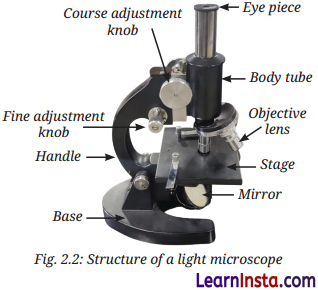
- A cell is usually too small to be seen by the unaided eye. The human eye has a limit of resolution of 0.1 mm. Two points closer than 0.1 mm appear as a single point when viewed from 25 cm. A convex lens or combination of lenses (objective lens and eyepiece) is used to magnify objects too small to be seen with the naked eye.
- Robert Hooke in 1665 was the first to observe cells using a self-designed microscope (200 – 300X magnification) while examining a thin slice of cork.
- Light microscope uses visible light and different objective lenses (10X, 40X) to achieve magnification and resolution.
- Total magnification is the magnifying power of eyepiece multiplied by the magnifying power of objective lens.
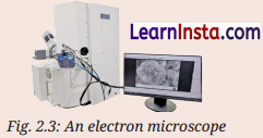
- Electron microscope uses a beam of electrons instead of light to produce highly magnified images at the nanometre scale.
- Over the years, scientists have improved the microscope by improving its three main features e.g. resolution (measure of clarify), contrast (the difference in brightness between various parts of an object), and magnification which resulted in making microscope a powerful tool for studying cells.
![]()
Structure of a Cell
Cells are organised into specialised tissues and organs, and collectively perform specific function. For these cells to function as units, they must be able to interact with one another and with their surroundings.
→ Cell Membrane – The Universal Feature of a Cell:
- Universal feature of a cell is its cell membrane (plasma membrane). It is a thin, selectively permeable boundary (7 – 10 nm thick) surrounding all cells. It allows some substances to pass through while blocking others.
- The movement of water through a selectively permeable membrane from a region of higher water concentration to a region of lower water concentration is called osmosis.
- Net movement of particles from higher to lower concentration, even without a membrane is called diffusion. Osmosis is the diffusion of water across a selectively permeable membrane. In plants, water from the soil enters root cells by the process of osmosis.
- In isotonic solution, the solute concentration outside the cell equals solute concentration inside the cell. In hypotonic solution, solute concentration outside the cell is less than inside the cell; water enters the cell. In hypertonic solution, solute concentration outside the cell is greater than inside the cell; water leaves the cell.
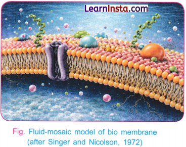
- Fluid-mosaic model explains the structure of the cell membrane as a lipid bilayer with embedded proteins which act like gatekeepers in helping substances pass through. The molecules can move, making it fluid. Protein molecules are arranged like tiles in a mosaic. Hence it is called mosaic model.
→ Cell Wall – The Outer Covering of Cells:
- All living cells communicate with their surroundings and their neighbouring cells through the cell membrane. However, cells of plant, fungi, and bacteria have an additional layer around the cell membrane, called the cell wall.
- The cell wall is an additional rigid covering outside the cell membrane found in plant cells, fungi, and bacteria. It is made mainly of cellulose in plants. It is permeable and provides shape, strength, and rigidity.
- Animal cells lack a cell wall, so they can change shape easily, supporting movement and tissue function.
![]()
The Cell Interior – A Coordinated Working System
Plasma membrane, cytoplasm, and nucleus are the three basic parts of most cells. Plasma membrane is selectively permeable membrane. Cytoplasm is a semi-fluid jelly like substance.
In addition to the prominent nucleus, the cytoplasm contains several sub-cellular components called organelles, along with other substances present in it, most of which are only visible with an electron microscope.
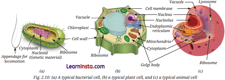
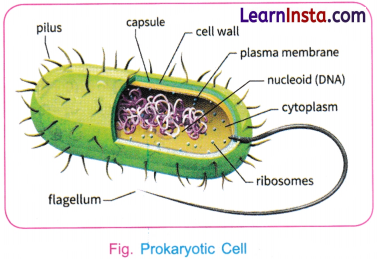
Prokaryotic (pro means primitive and karyon means nucleus) cells lack a well-defined nucleus and membrane-bound organelles. Example: bacteria.
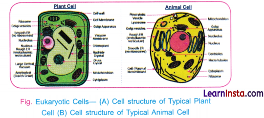
Eukaryotic (eu means true, and karyon means nucleus) cells have a well-defined nucleus and membrane-bound organelles. Example: plant and animal cells. In eukaryotic cells, a network of fine fibres forms the cytoskeleton, which provides structural support, maintains cell shape, and enables cell movement and internal transport. Eukaryotic cells carry out various life processes in different cell organelles independently at the same time.
Viruses are a cellular (no cells) infectious agents that are too small to be seen under a light microscope. Viruses are composed of some genetic material with a protein coat.
![]()
Cell organelles help in building new materials, removing waste, and providing energy to the cell. They work together to perform all functions of a cell. Thus, a cell is like a tiny living factory, with each of its parts performing a specific job.
Why do Eukaryotic Cells need these Organelles?
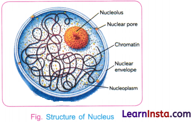
Nucleus has a double-layered nuclear membrane with pores. It contains chromosomes (DNA+ proteins). DNA carries genetic information. Functional segments of DNA are called genes.
Some cells are specialised to perform specific functions. For example, mature Red Blood Cells (RBCs) in humans do not have a nucleus (enucleate). The absence of a nucleus provides more space for haemoglobin, allowing it to transport a larger amount of oxygen to all cells of the body. Since they lack a nucleus, they cannot repair or divide themselves. As a result, their lifespan is short and they survive approximately for 120 days. The sieve cells in phloem are also enucleated.
Prokaryotic cells do not have a well-defined nucleus. Their DNA is present as a single circular molecule associated with specific proteins. The region containing this genetic material is called the nucleoid.
Ribosomes are the tiny structures that are sites of protein synthesis, found freely in cytoplasm or attached to the Endoplasmic Reticulum (ER).
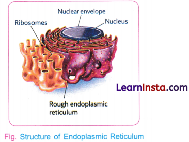
Endoplasmic Reticulum (ER) is a large organelle which spreads like a network within the cytoplasm. Rough Endoplasmic Reticulum has ribosomes (protein synthesis) while Smooth Endoplasmic Reticulum lacks ribosomes (lipid and hormone synthesis).
![]()
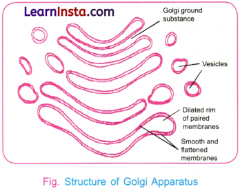
Golgi apparatus has stacks of flattened sacs that modify, sort, and help in package of proteins and lipids for transport, secretion, or lysosome formation. It acts like the cell’s post office.
Lysosomes are single membrane-bound sacs filled with enzymes that break down waste materials and damaged organelles. Human sperm cells contain lysosomal enzymes. When a sperm meets an egg, these enzymes help break down the outer layer of the egg, allowing fertilisation to take place.
Mitochondria are double-membrane organelle; the inner membrane has folds called cristae which increase the surface area for chemical reaction and facilitate energy production. Mitochondria break down glucose to release energy that is stored as ATP (energy currency of the cell). These are called the powerhouse of the cell.

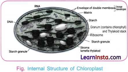
Plastids are found in plant cells. These are of three types: Chloroplasts are green in colour and help in photosynthesis, Chromoplasts are coloured and attract pollinators, Leucoplasts are colourless and store food.
Inside the chloroplasts, there is a semi-fluid substance matrix, called the stroma. Stroma are disc-shaped membrane structures that contain chlorophyll which absorbs sunlight during photosynthesis. Stroma stores the sugar synthesised during photosynthesis.
Mitochondria and plastids have some features that are similar to certain bacteria. For example, they both have their own DNA and ribosomes, and thus, they can make some of their own proteins. These characteristics suggest that both mitochondria and plastids share an evolutionary history with these single-celled organisms.
In a mature plant cell, there is one large central vacuole which is filled with watery fluid called cell sap. The membrane of the central vacuole is called tonoplast. The vacuole stores water, minerals, sugar and waste material and thus helps maintain pressure inside the cell, thereby keeping the plant cell firm (turgid). When a plant does not get enough water the vacuole loses water, the cells become less firm, and the plant gets wilted. In animal cells vacuoles are small.
![]()
How do Normal Cells Grow and Divide
- In 2010, scientist J. Craig Venter and his team made an important discovery in the field of synthetic biology. They first studied the complete DNA sequence of a simple bacterium called Mycoplasma mycoides using computer programming. Then, they chemically synthesised (in the laboratory) an exact copy of this DNA. They also performed an experiment to show that DNA controls the structure and activities of the cell.
- Cells in our body can grow and divide to replace the old, dead, or damaged cells. Process by which new cells are formed from pre-existing cells is called Cell division. It is important for growth, repair, and reproduction.
- Everyday, an estimated hundreds of billions of cells in our body are replaced, which is almost 1 per cent of the total number of cells in our body. Both prokaryotic and eukaryotic cells divide, but eukaryotic cells divide in a more controlled and orderly manner by a process called the cell cycle. Cell cycle is the controlled and orderly process by which eukaryotic cells divide.
Cell Division
- There are two major types of cell division, mitosis and meiosis. Mitosis is important for normal growth, repair, maintenance and asexual reproduction, while meiosis is important for sexual reproduction for creation of genetic diversity.
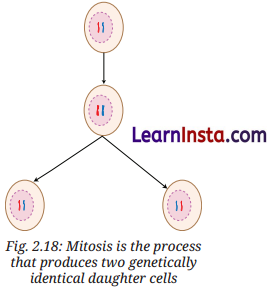
- Mitosis produces two genetically identical daughter cells from one parent cell with same number of chromosomes as the parent cell. It is important for growth, repair, and asexual reproduction.
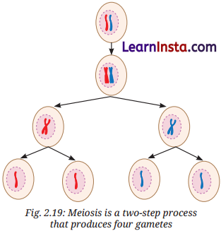
- Meiosis is a type of cell division that produces gametes and occurs only in the cells of reproductive organs. Gametes produced for sexual reproduction create variations and diversity among living organisms. Therefore, children resemble their parents but are not exactly the same.
- Meiosis occurs only in reproductive organs. Parent cell divides twice to produce four daughter cells, each with half the number of chromosomes. It is important for sexual reproduction and genetic diversity.
- Scientists have developed methods to grow plant and animal cells outside the body in special conditions. This is called cell culture. Cell culture is crucial for studying how cells work and for the production of biochemicals, food, medicines, vaccines, and more.
- The processes of mitosis and meiosis must occur in a proper and controlled manner. If there is any error in these processes, it can lead to various problems in the body of an organism like uncontrolled cell division, tumour formation, and abnormal chromosome number.
- Errors in meiosis may result in genetic disorders, developmental problems, early pregnancy loss, or reduced fertility.
![]()
Cell Theory – The Unifying Principle of Biology
All living organisms are made up of cells. In 1838, a German botanist named Matthias Schleiden reported that all plants are made up of cells. In 1839, German zoologist Theodor Schwann found that all animals are also made up of cells. Later, in 1855, a German scientist named Rudolf Virchow further expanded the Cell Theory by stating that new cells are formed only from pre-existing cells. Together, their work led to the formulation of the Cell Theory.
→ Classical Cell Theory states:
- All living organisms are made up of one or more cells.
- The cell is the basic unit of structure and function.
- All cells arise from pre-existing cells.
Do Cells Grow and Reproduce Forever?
Cells grow and divide in a controlled way, stay in the right place, carry out their functions, and eventually die when they are no longer needed. Dead cells are replaced by new cells that carry out the same function. In many animal cells, cell division stops when cells come in contact with neighbouring cells. This is called Contact inhibition. Cancer cells lose this control and result in formation of tumours.
Normal cells grow, age and die in a controlled manner. Sometimes, this system breaks down, and abnormal cells start growing and dividing uncontrollably. This results in the formation of tumours, which may be benign or malignant. Cancerous tumours can invade nearby tissues and even spread to other parts of the body to form new tumours.
Cells also have natural ways of dying to maintain a balance. Programmed Cell Death (PCD) is a genetically regulated and organised process of selective cell destruction. It is essential for normal development, cellular quality control and immune function.
Plant cells grow differently. Due to their rigid cell walls plant cells do not show contact inhibition and follow a different pattern of growth.
The ability of a plant cell to develop into a complete plant if provided with suitable nutrients and conditions is called Totipotency. It was proposed by Gottlieb Haberlandt (1902). His idea laid the foundation for Plant Tissue-culture Technology. Growing cells outside the body in nutrient-rich media under controlled conditions is called Cell culture.